

by Taylor Howard, DVM
Veterinary diagnostics are integral in our approach toward understanding your pet’s overall condition. While nothing can replace a thorough physical exam, imaging modalities are often required for a complete picture of what is going on internally, as well as externally. Whether the veterinary visit is for a routine annual wellness exam or something more specific, some of these modalities may be offered. As with all tests, none of these are perfect for every situation. An understanding of what these tests are and how they work will help to guide our decision making and planning process. The following is a brief description of those most commonly used in today’s practice.
Radiology/X-ray
These are two dimensional images made by passing radiation through a focused point on the body. The image is captured traditionally by the use of film, similar to how photographs are made. The areas of the film that capture radiation turn black. The denser the tissue is, the less radiation can pass through. Bones (dense material) will appear white, while organs and air (less dense material) will appear black. Soft tissue structures such as muscle and intestines will vary somewhere in between, providing a level of grey.
In recent years, film has been gradually replaced with digital recording devices. The principles are similar in both cases. X-rays are used to evaluate bones for fracture, the size and shape of organs, identify some foreign objects in the intestines or stones in the bladder.
The use of X-ray imaging is the most common of all imaging modalities because of the availability in every practice and it is also the most affordable imaging test. For this reason, it is often required to be performed prior to any of the other imaging options available.
Contrast Studies
This is a subcategory of X-ray imaging used for outlining or highlighting structures of interest. The contrast medium is a liquid of some type that absorbs all radiation, similar to dense objects and will show up appearing white like bone.
The most commonly used contrast is barium. Barium given orally or by enema helps to outline the intestinal contents. This is helpful when a foreign object is suspected but cannot be seen with a routine X-ray because the object is often times the same density as the surrounding tissue and cannot be seen otherwise. This is usually done as a series of images over time to evaluate the progression of contents through the GI tract.
Contrast can be administered as an injection into the bloodstream to see how the kidneys are functioning in patients with kidney failure, and into the spinal column to isolate a herniated disc. These techniques as well as others, are available and can be used to capture a more detailed and localized snapshot of your pet’s health.
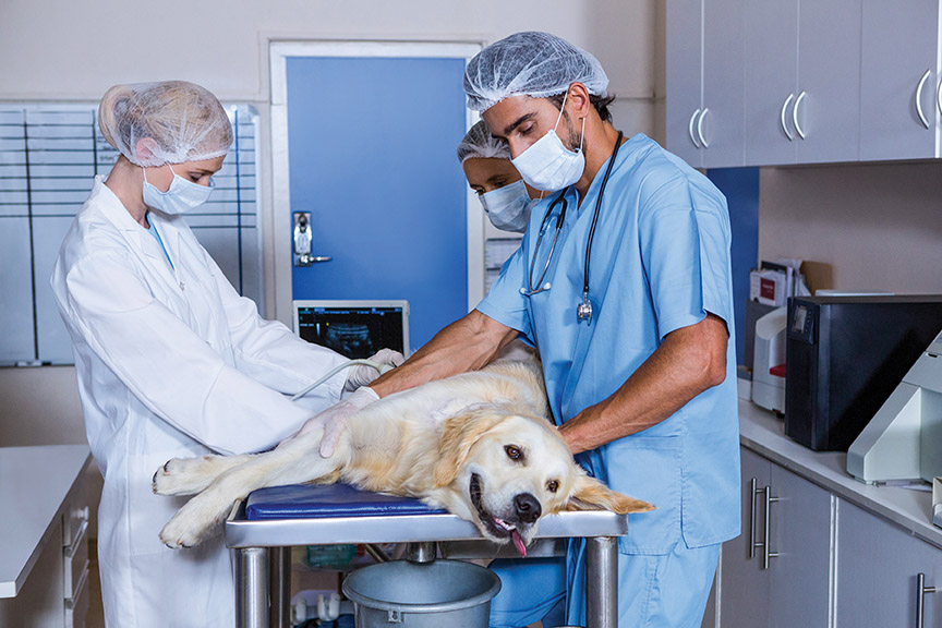
Ultrasound
Another name for ultrasound is sonogram. Unlike X-rays, ultrasounds use no radiation. An ultrasound uses sound waves emitted from a wand to bounce against tissues. These sound waves are either absorbed in the tissues, travel all the way through the tissues, or are reflected back. The wand also acts as a sensor to pick up the sound waves as they come back like an echo. The wand requires direct contact with the skin, so the fur is shaved in most cases where the focus is concentrated. Ultrasound is painless and does not usually require anesthesia or sedation.
Ultrasound penetrates the tissue on a single plane showing, in essence, a slice through every layer revealing minute architectural detail. The detail attained can then be interpreted to sufficiently describe a suspected lesion located with a previous X-ray.
Ultrasound imaging looks closely at a specific site whereas radiology is used to see the big picture. It is understandable that if an ultrasound is used prior to X-ray imaging some things may be missed. This is why X-rays are taken prior to an ultrasound in most cases.
Ultrasounds can be used in suspected heart disease cases. An X-ray is used to see an outline of the entire chest cavity to see what changes or abnormalities may be occurring. Then an ultrasound is used to look directly at the heart. The ultrasound captures more refined details including measurements of blood flow, muscle thickness, valve integrity and so on. This is called an echocardiogram and is essential for a definitive diagnosis and a therapeutic plan.
There have been some recent advancements in the use of ultrasonography in chemotherapy. The tumor is visualized and a catheter is inserted while watching the image, targeting areas where a chemotherapy agent is injected directly. This targeting approach reduces risk by avoiding systemic circulation from intravenous administration.
One major limitation is that ultrasound does not reflect when traveling through air. Therefore, lung tissue cannot be evaluated by way of ultrasonography. In the event that a lesion is suspected in the lungs, CT imaging is sought.
Computer Tomography (CT)
Another common name for this type of imagery is CAT Scan. This is a more advanced type of radiology that has been used for many years in human medicine. A series of X-ray images are taken in sections. The pet must remain still and for this reason anesthesia is required. Each section is advanced forward or backward over a varying period of time based on the size of the area. A mouse would take much less time than a Great Dane for example. The images are then reassembled into a three dimensional map that can be navigated through an advanced software program. A contrast medium can be used like with regular X-rays to enhance or highlight these structures. A CT image can find minute abnormalities and is used commonly when planning a surgical approach in a very sensitive area. Examples are spinal cord injuries, complicated fracture repairs and tumor removals with important vessels or structures within.
Utilization of this type of imaging is becoming more available as the cost of equipment becomes more feasible. Human hospitals upgrade their CT technology frequently, thus making the previous equipment available at reduced price.
Magnetic Resonance Imaging (MRI)
As with CT imaging, pets are anesthetized while advanced equipment takes a series of images over time and reassembles these images into a three dimensional format for viewing by a specialist. This technology uses magnets and radio waves instead of X-rays, making it safer. It is more sensitive to small structures down to less than a millimeter in size. It is reserved for scanning neurological structures primarily. Spinal cord or brain injuries can lead to severe problems when something as small as a blood clot (i.e. aneurysm) is all that stands in the way of recovery.
The cost of equipment limits its use in the general practice. Regardless of expense, specialized hospitals with board certified veterinary radiologists, internists, orthopedic surgeons and neurologists are generally within reach. Referrals are made in the event that an MRI is needed.
Dr. Taylor Howard attended Utah State University and graduated from Ross University with his Doctor of Veterinary Medicine. He worked in mixed animal practice, emergency medicine, general practice, and dentistry in Oregon before moving back to Utah, where he joined University Veterinary Hospital & Diagnostic Center. His goals in practice are toward diversity in exploring medicine, communicating with his clients and being part of a major cornerstone in his community.


